histology facility
The TRI Histology Core Facility was established in 2014 and is located on level 5 North East wing, in rooms 5029 and 5031. It is a tissue preparation facility enabling demonstration of morphology through brightfield or fluorescent microscopy, and other downstream analysis options. Our aim is to provide efficient, high quality flexible services with responsive turn-around time to researchers within TRI as well as external clients. We also provide training and access to a range of specialised equipment.
For more information regarding fees, please refer to PPMS, or contact the TRI Histology Core Facility staff.
|
what can we do? |
|||||
ProcessingStandard processing |
EmbeddingStandard embedding |
sectioningFrozen tissue sectioning |
labellingSlide labelling |
immunostainingIHC, IF, ISH and Multiplex |
histochemicalAlcian Blue + PAS |
|
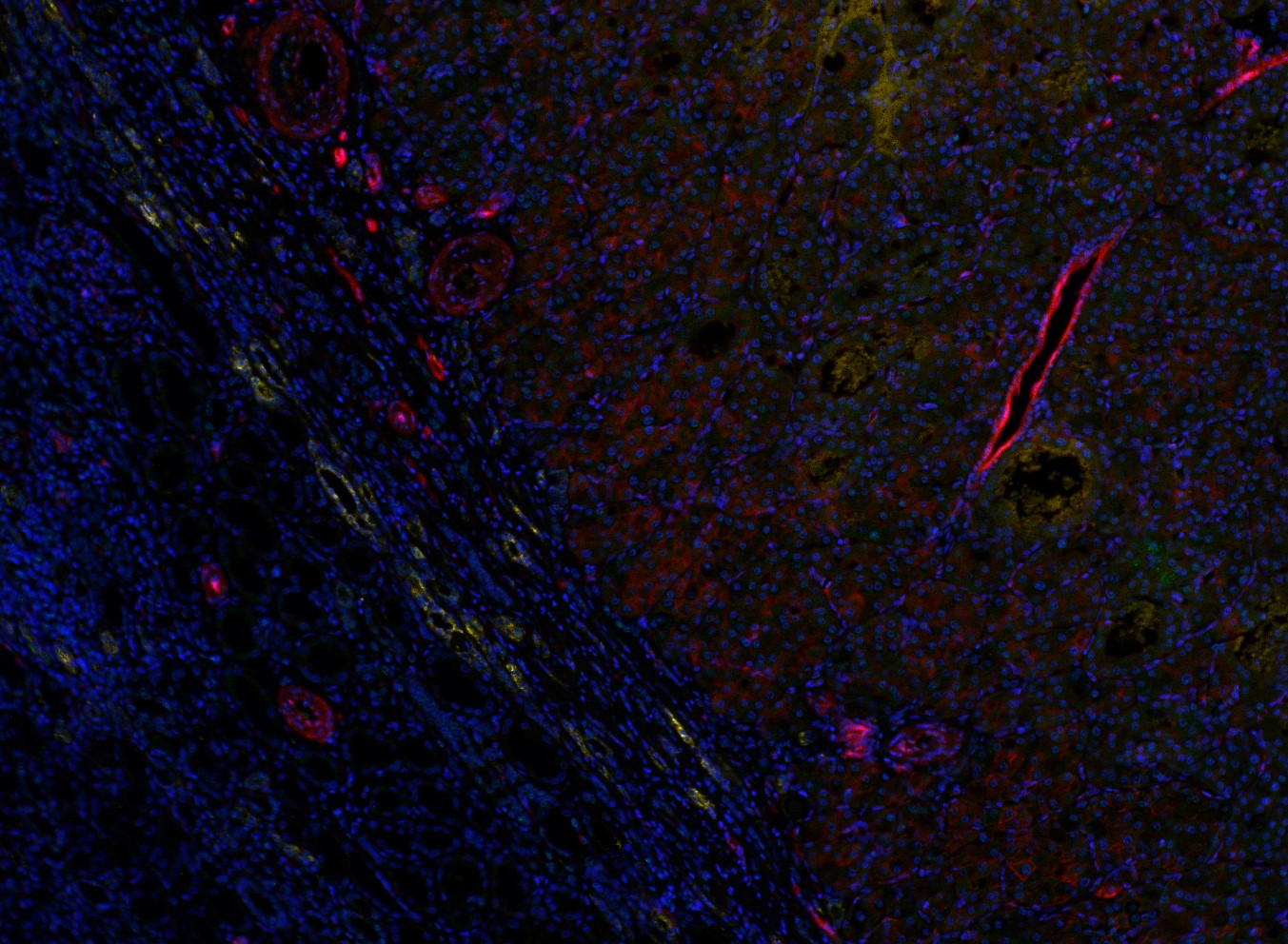
what is multiplex staining?
|
|
||||
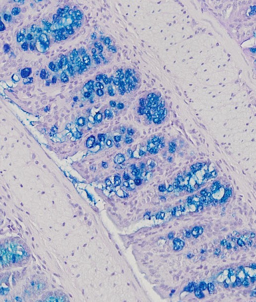 Colon stained with Alcian Blue |
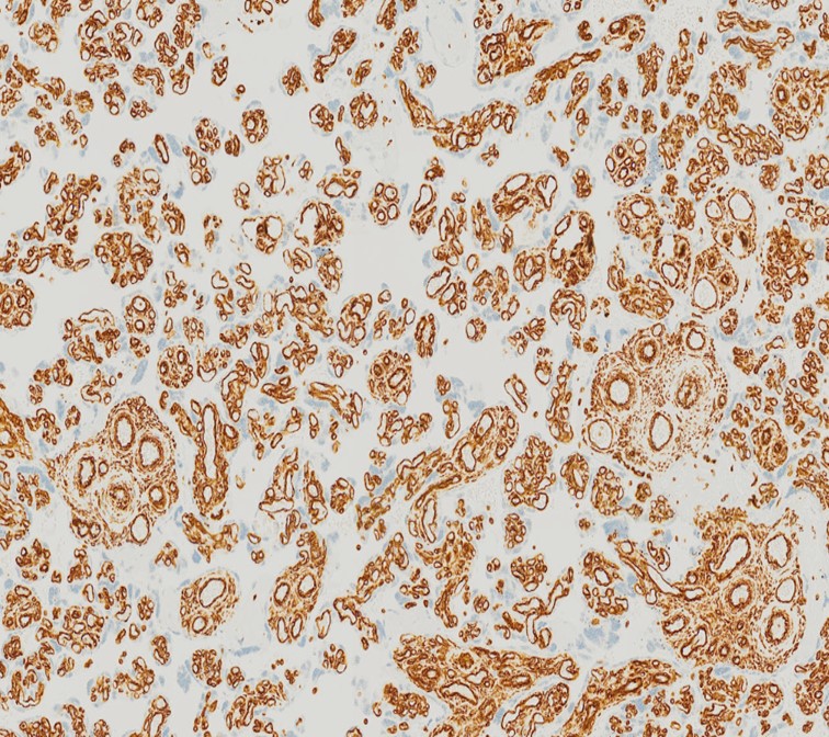 Placenta stained using IHC |
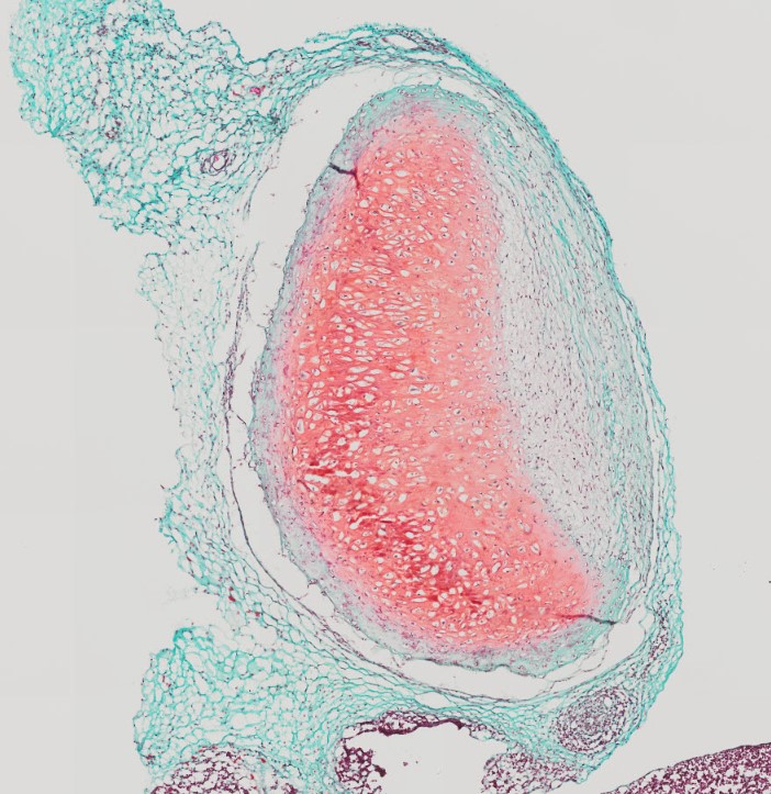 Cartilage stained with Safranin O |
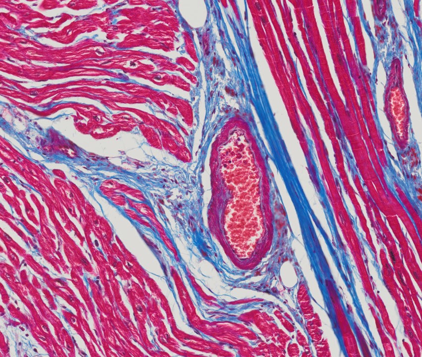 Heart tissue stained with Masson Trichrome |
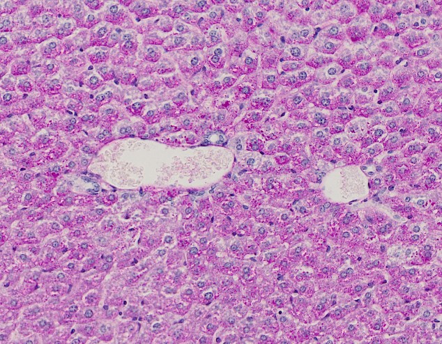 Liver stained with PAS |
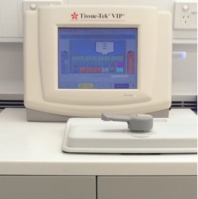 |
tissue processor - vip6 Designed for the automated dehydration and paraffin infiltration of human, plant and animal tissues. Fixed samples are submerged in a series of increasing concentrations of ethanol, then several changes of xylene, and finally infiltrated with paraffin prior to embedding. Protocols can be customised to the sample's needs with variable times, temperatures, and pressure and vacuum cycles. Contact the Histology Facility Staff for more information. |
 |
embedding station Following tissue processing, this station is used to position and embed tissues in molten paraffin wax to form paraffin blocks ready for microtome sectioning. |
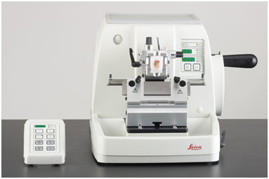 |
microtomes We have two rotary microtomes designed to slice formalin fixed, paraffin embedded tissues into sections as thin as 1μm. These sections are then picked up on microscope slides and used in a range of staining applications, or even protein extraction. |
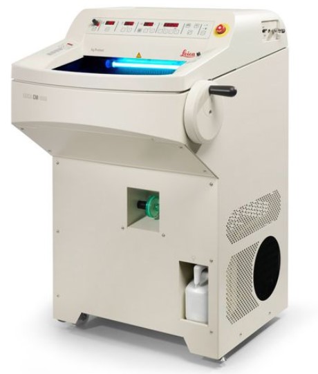 |
cryostatS The cryostats are used for sectioning fresh or fixed tissue samples, frozen with OCT embedding medium, to provide rigidity and support while sectioning. Fresh bone samples can be sectioned using the tungsten blade on the Leica CM1950. The ability to section unfixed samples is useful for fat studies, and investigating temperature or fixation senstive antigens. |
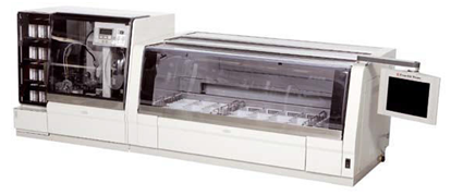 |
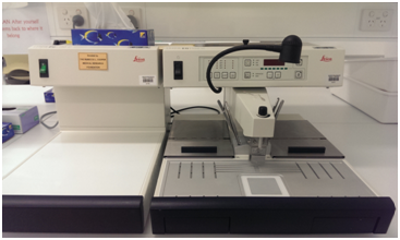
tissue-tek automated stainer and coverslipper For all of your routine and special histochemical stains, the Histology Core Facility has a programmable automated stainer to produce consistently high quality stained slides. It is linked to an automated coverslipper which will then transform your slides into clean coverslipped slides ready for imaging in Microscopy. The coverslipper can be used independently of the stainer, so why not bring your slides prepared at the bench for superior coverslipping. |
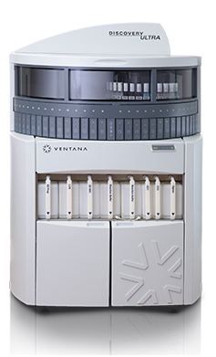 |
ventana discovery ultra The Discovery Ultra enables high quality, highly reproducible chromogenic and tyramide flourescent immunostaining. It performs all operations required to automatically process slides for immunohistochemistry (IHC) and in situ hybridisation (ISH) staining. Capabilities include: Baking, deparaffinisation, antigen retrival and washing can be customised. Single or multiplexed chromogenic and TSA flourescent IHC, ISH and IF staining. It has 30 independent slide reaction chambers with individual slide heaters. |
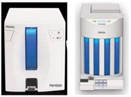 |
slide and cassette printers The slide and cassette printers produce highly legible labels to clearly identify your samples, and minimise transcription errors in subsequent steps of processing and analysis. |
CHARGES and terms of use
To enquire about facility charges please email [email protected] or access the PPMS booking system.
TRAINING
The TRI Histology Facility offers a monthly Introduction to Histology course, targeted at users new to the field and those who would like a refresher. Please contact Histology Core Facility Staff for session times.
Access to the facility
Before using the facility, researchers and students must undertake one on one equipment specific safety inductions and training. Laboratory staff will supervise new users and determine when users are competent to undertake independent use.
Access for new users is from 8am - 4pm Monday to Friday. After hours use may be requested, and granted after a review of usage and with approval by the users supervisor.
All users must book equipment prior to use through PPMS. For any queries regarding this system, please contact [email protected]
To find out more about the equipment and services, or to book for an induction and training, contact the facility on ext. 37065 or email [email protected]


